Ksena | micro Longrin | world
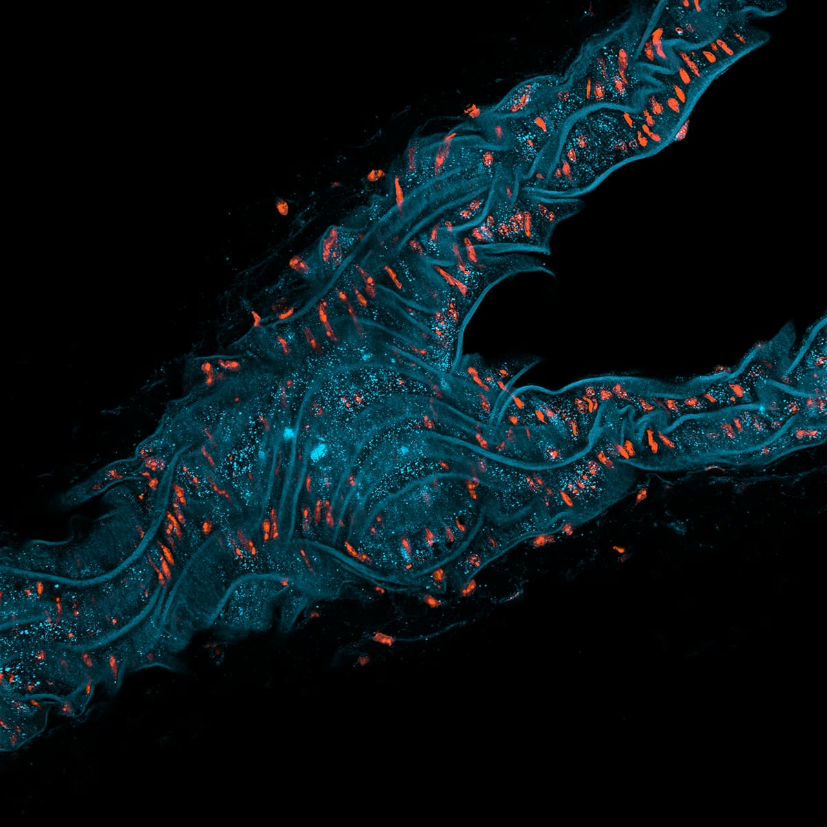
Mouse lung cross section
Interestingly oriented cross-cut muscular tubular structure- either a tiny airway or an arteriole. You can see the long elastin fibres wiggling longitudinally in the plane of the wall, and bands of smooth muscle nuclei arranged circumferentially inbetween elastin layers.
🟦Autofluorescence 🟧 DNA
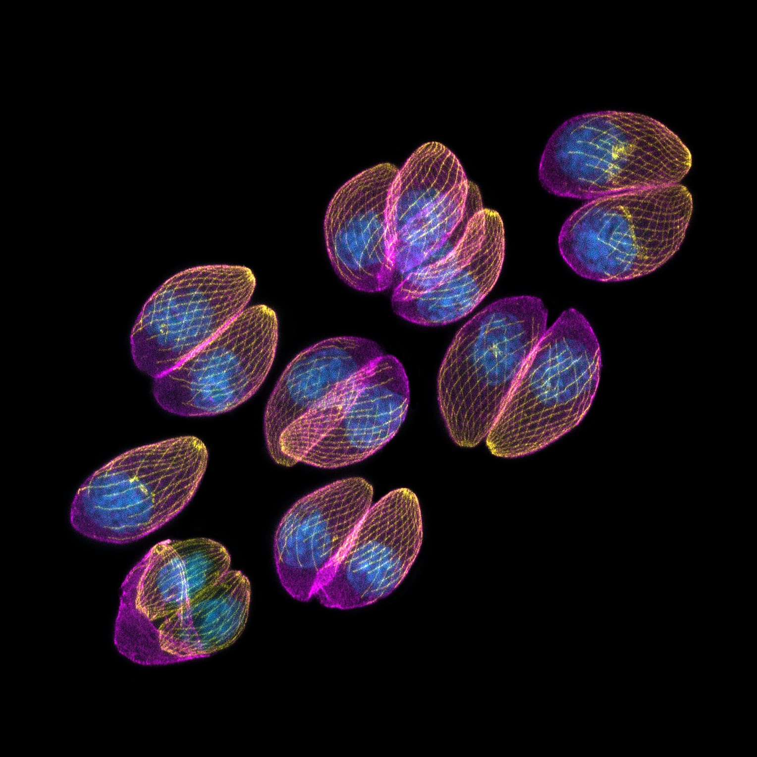
Toxoplasma parasites in human skin cells
Winner — Nikon Small World 2024
Acute-stage Toxoplasma gondii parasites in a human skin cell, showing the inner membrane complex (magenta), microtubules (yellow), and nuclei (blue). Eight parasites originally infected this fibroblast; six have already divided once or twice via endodyogeny, and the bottom parasite is caught mid-division, with two daughters emerging from the remnants of the mother's shell.
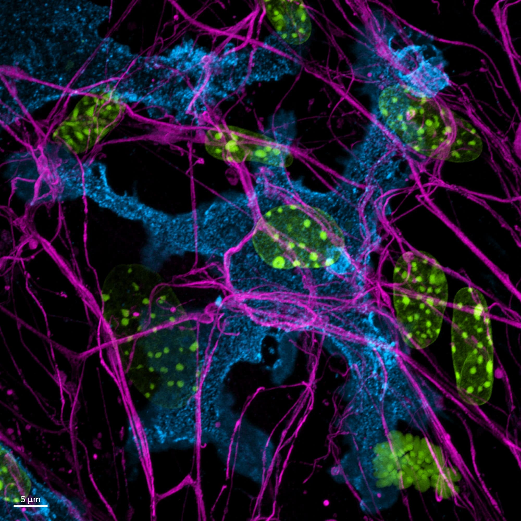
Macrophage patrolling nerve cell culture
This is the dissociated culture of mouse dorsal root ganglia neurons, where immune cells like this macrophage sneaked in past our filters.
I find macrophages somewhat disturbing and mildly threatening. I mean, check this one. It looks like a cyclops ready to devour you whole, just as they did back when Greece was ancient and full of wonders.
🟥Nerves 🟦Macrophages 🟩DNA
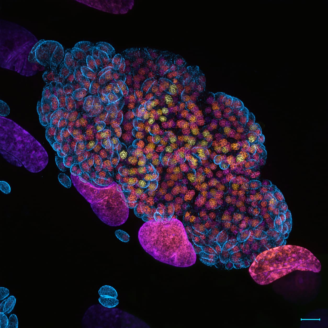
Acute-stage Toxoplasma parasites living in human skin cells
Winner — Huygens Image Contest 2024
Acute-stage Toxoplasma parasites, overgrown after three days of infection of human skin cells.
The DNA of every cell is depth-coded, so colour tells you how deep in the sample a nucleus sits — the higher up, the warmer.
The larger nuclei belong to the skin cells, the small tightly packed ones to the parasites.
🟦 Parasite inner shell (GAP45) 🟨🟧🟥🟪 DNA (depth-coded)

Purkinje neurons in mouse cerebellum
A slice through a mouse cerebellum, showing Purkinje neurons — the cells behind balance and coordination. They normally sit in one neat row, but a diagonal cut through the fold caught several rows at once, hence the splash. Their red glow comes from Kv3.3, a protein that lets these cells fire fast enough for precise movement. In people, a faulty Kv3.3 causes spinocerebellar ataxia type 13, a disease that damages and kills Purkinje neurons.
🟥 Purkinje neurons (Kv3.3) 🟦 DNA
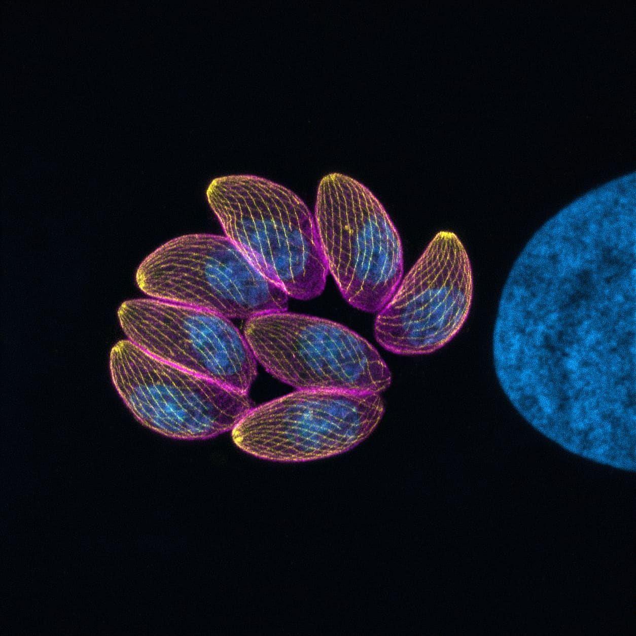
Acute-stage Toxoplasma parasites in a human skin cell
These eight Toxoplasma parasites were captured in the acute stage of infection, nestled inside a human skin cell (not visible here). The large blue structure on the right is the host cell’s nucleus. Notice the bright yellow dot atop each parasite - it’s the parasite’s “mouth”- used to invade the cell and deliver molecular tools that hijack and suppress the cell’s defenses.
🟪 Inner parasite shell 🟨 Parasite skeleton (microtubules) 🟦 DNA
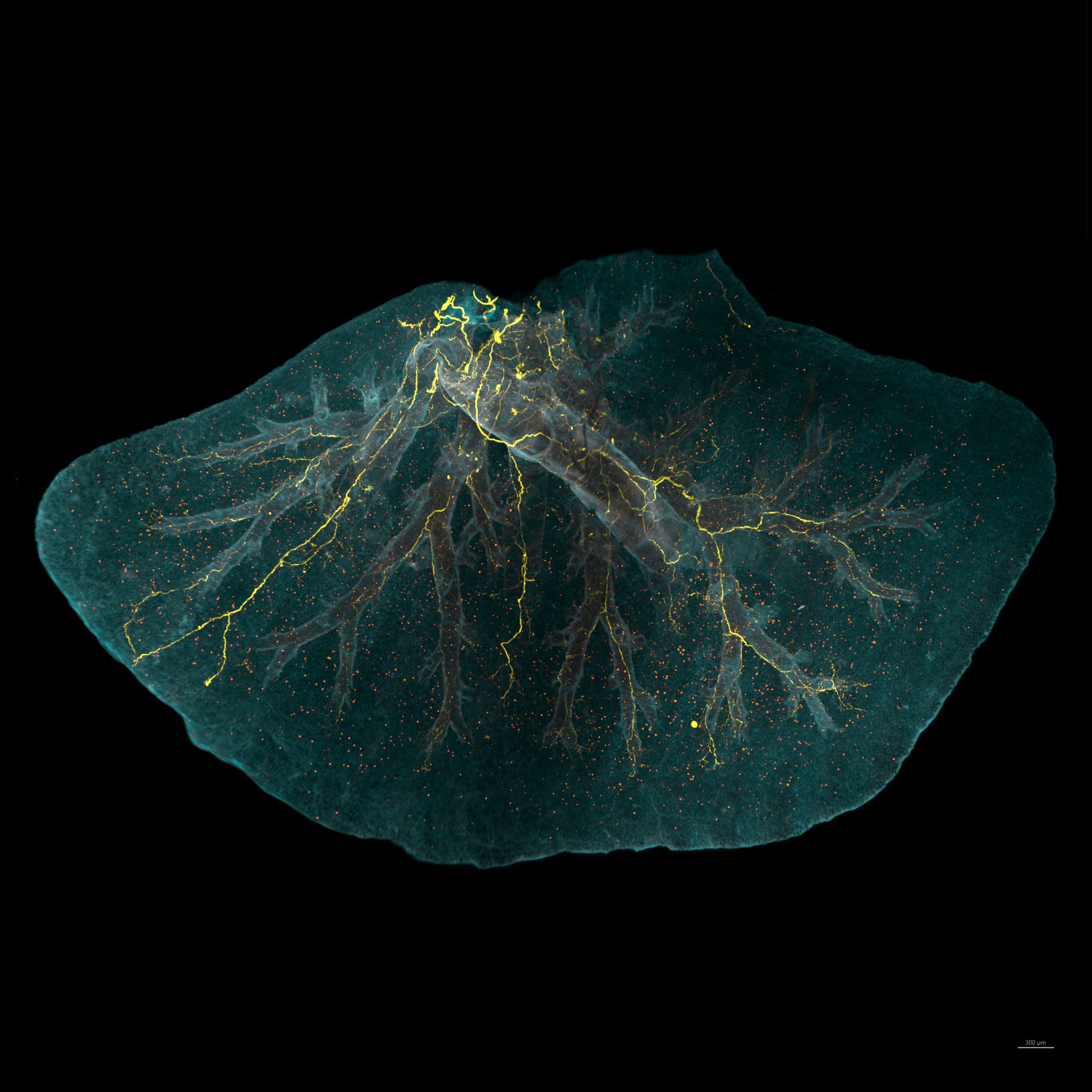
Mouse lung with transferred immune cells from another mouse
What counts as sci-fi to you?
For me, lately, is taking immune cells out of one mouse, dyeing them so they glow, putting them into another mouse - and finding them a week later, quite happily settled all over the new host's lungs.
🟨 Nerves 🟧 The transplanted immune cells 🟦 Autofluorescence
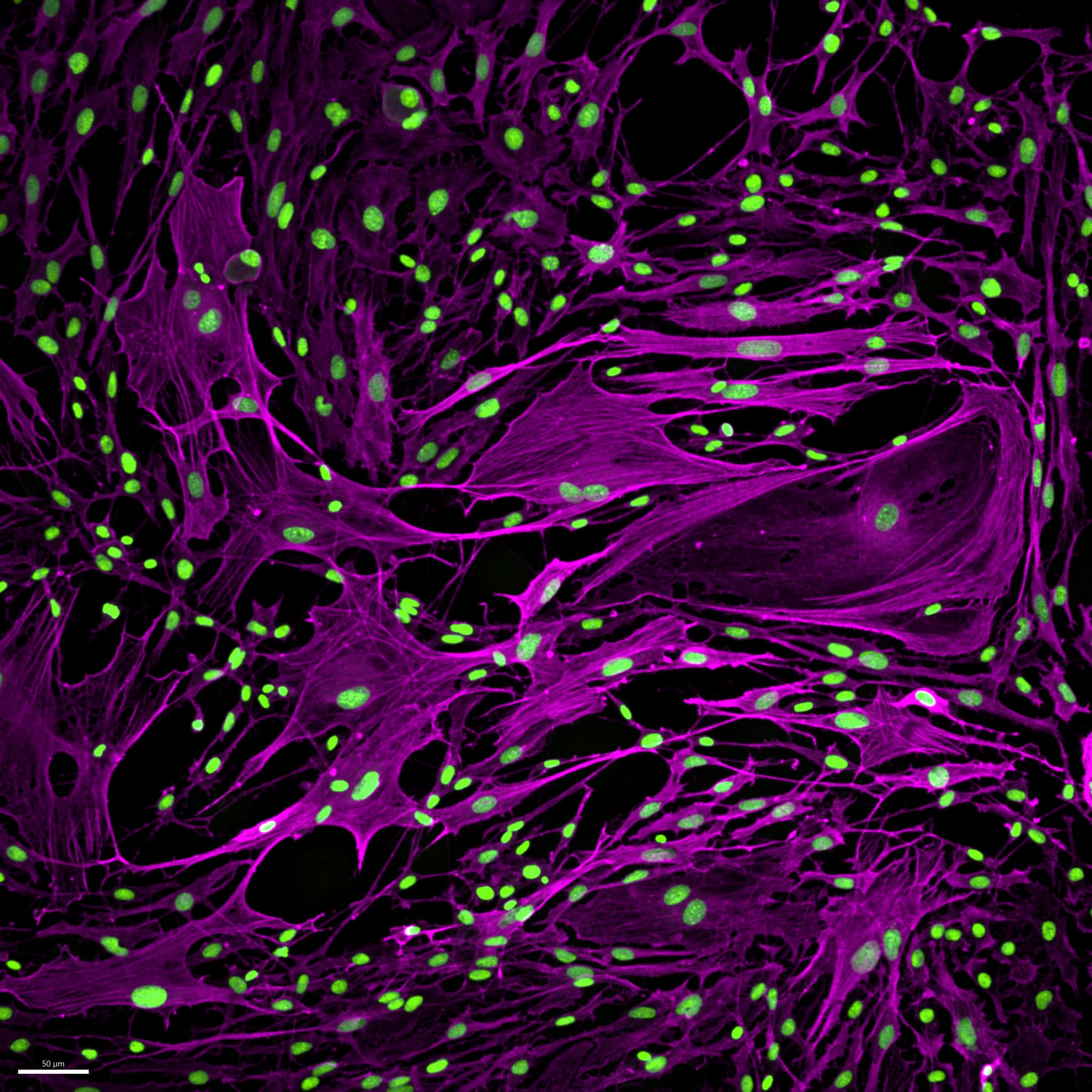
Neuronal cell culture
Sensory neurons from a mouse, teased apart and grown in a dish. They come from the dorsal root ganglia — the clusters beside the spinal cord that carry touch, pain and temperature signals from the body up to the brain.
🟩 DNA 🟪 Cell cytoskeleton (actin)
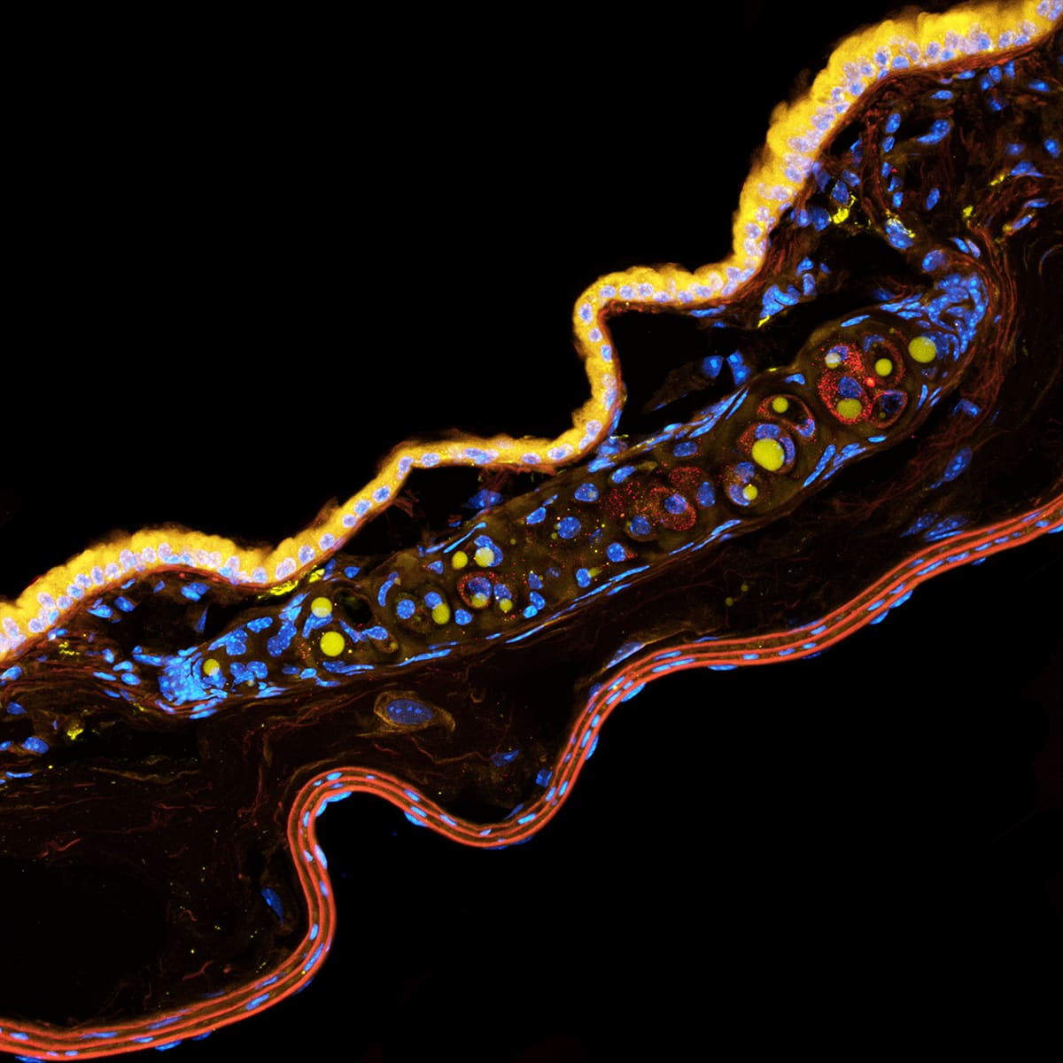
Mouse lung mucus-producing gland
A lengthwise slice through a mouse airway (a bronchus or bronchiole).
It shows three layers: the inner lining of cells (which have tiny hairs called cilia) coated in mucus that glows brightly under the microscope, glands beneath that produce the mucus, and a layer of muscle around the outside.
The gap you can see between these layers is probably just an artefact from how the tissue was prepared (from fixing or cutting it, causing it to shrink back), rather than a sign of any real disease or abnormality.
🟦 DNA 🟨 Non-specific stain 🟥 Non-specific stain
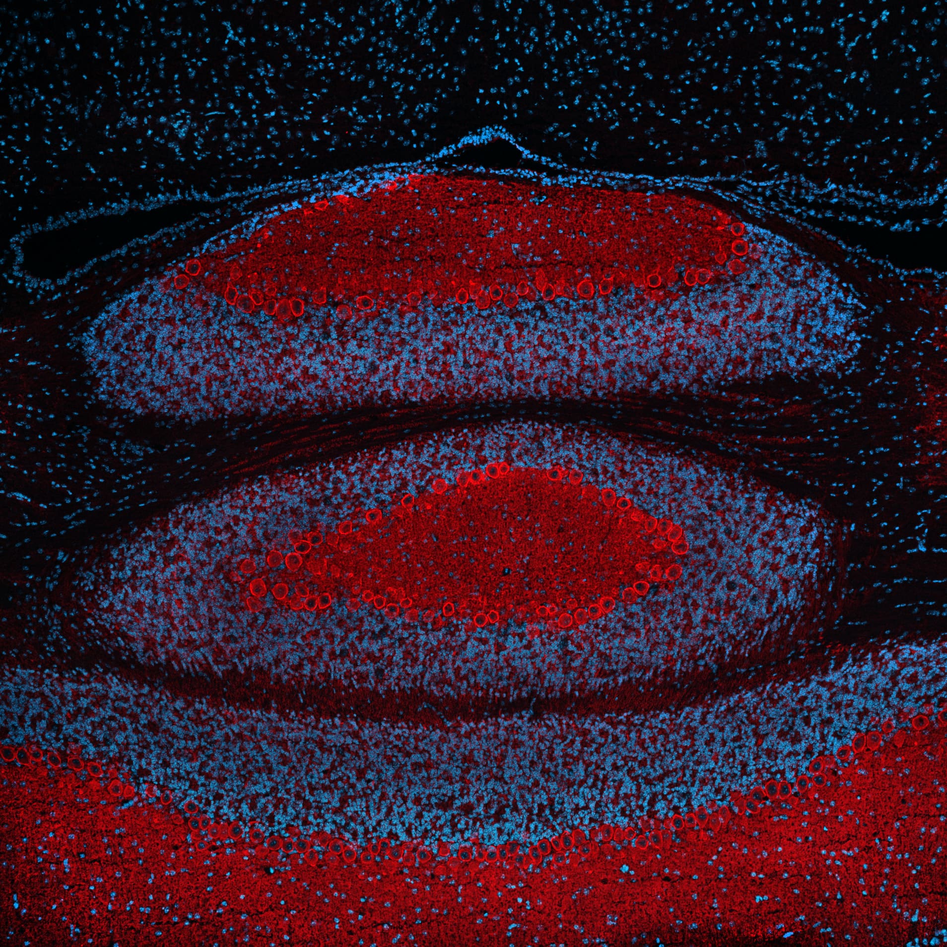
A crossection of a mouse cerebellum
A slice through a mouse cerebellum, the part of the brain that keeps you upright. The big red rings are Purkinje neurons, lined up single-file along the folds - the cells behind balance and coordination, the reason you can walk downstairs without thinking about it. Their red glow comes from Kv3.3, a protein that acts like a fast shutter on the cell membrane, cutting each electrical pulse short so the neuron can fire again straight away. When Kv3.3 is mutated, these cells sicken and die, causing an inherited condition called spinocerebellar ataxia type 13. I took this image while hunting for other mutations that might do the same.
🟦 DNA 🟥 Voltage-gated potassium ion channels (Kv3.3)
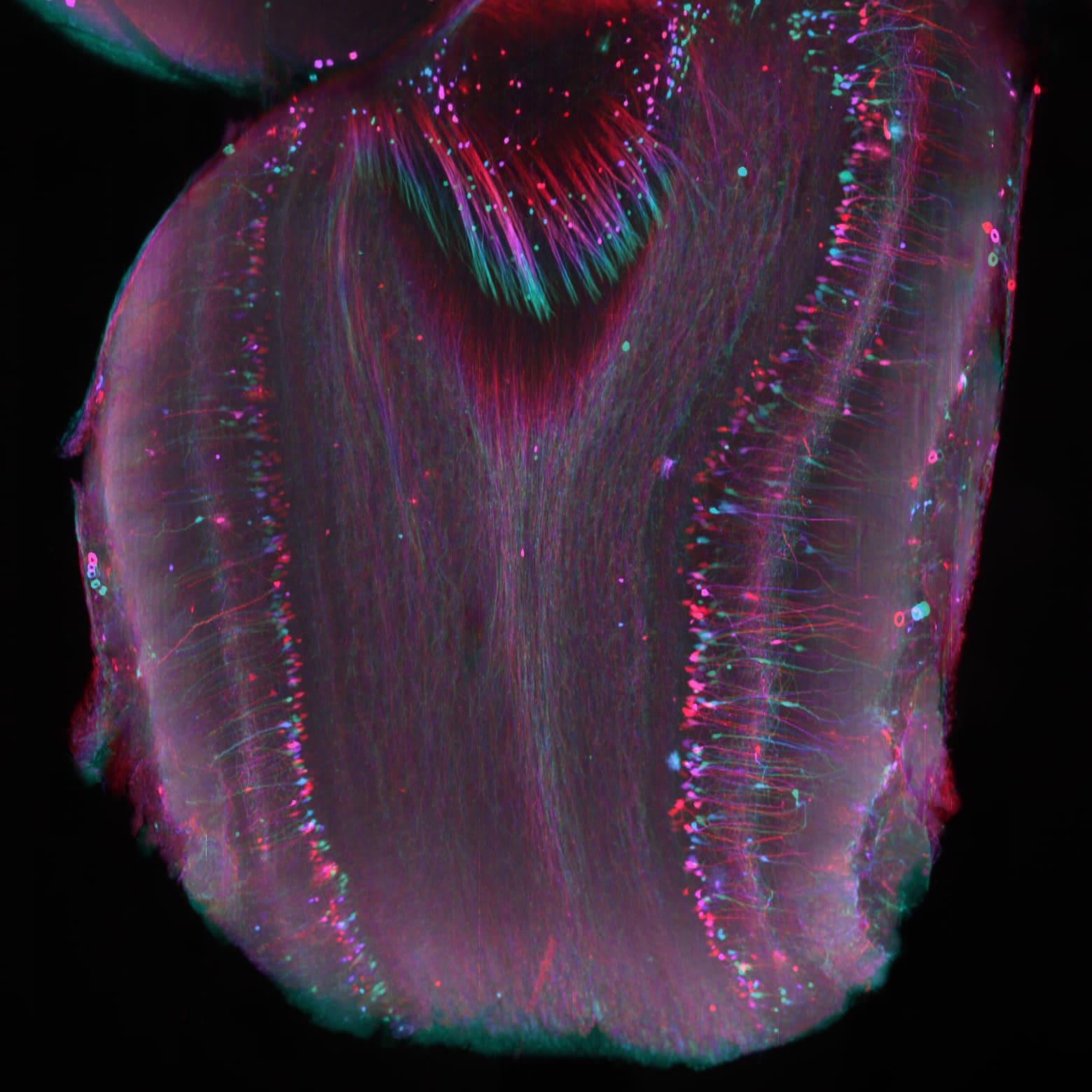
Mouse brain olfactory bulb crossection
A crossection of the middle of the mouse olfactory bulb, depth-coded.
The neurons you see express tdTomato fluorescent protein, with the signal boosted by an antibody.
The two cheerful rows of layered neurons are mitral cells.
When a mitral cell fires, it wakes its inhibitory neighbours, which quiet both it and the cells nearby. That contrast-sharpening is what makes two similar smells easier to tell apart.
🌈TRPV1-tdTomato (Transient Receptor Potential Vanilloid 1 tagged with tdTomato)
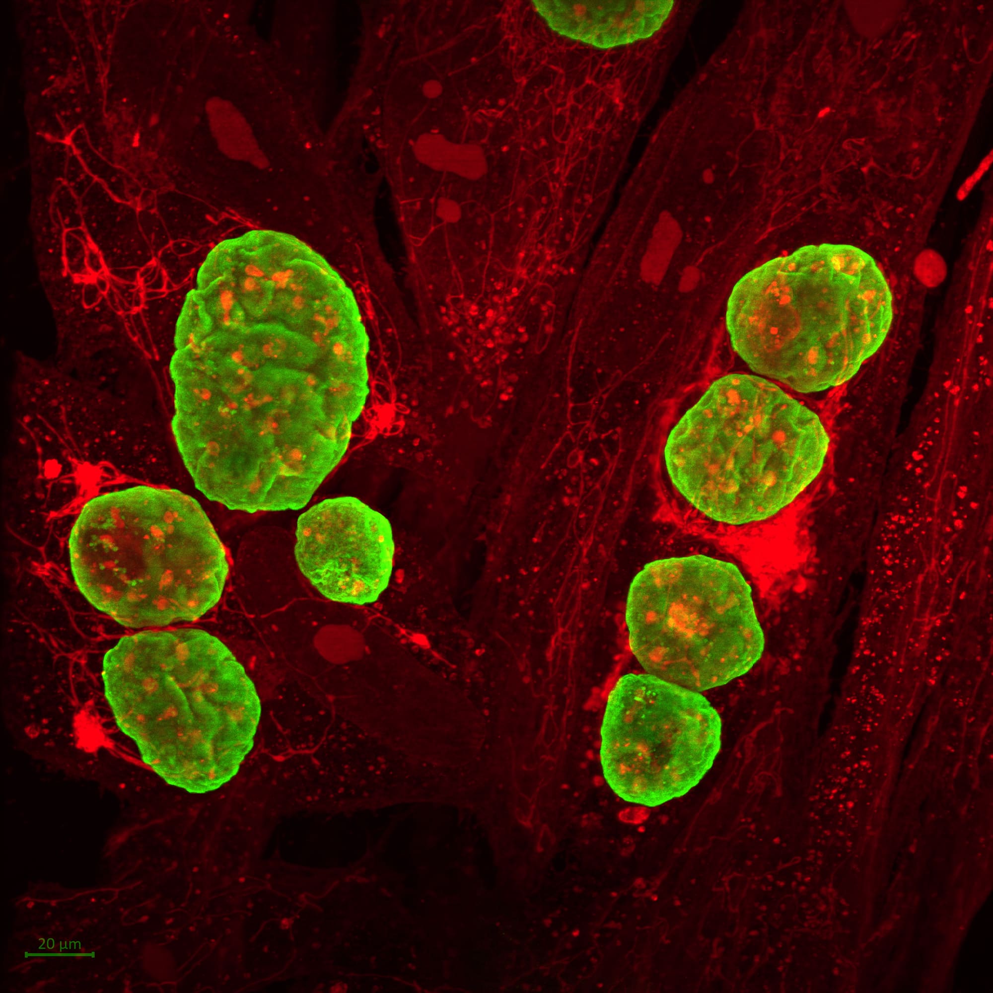
Chronic-stage Toxoplasma infection of a human skin cell
A human skin cell with an uninvited Toxoplasma guest.
The red web is the cell's own protein architecture, crisp and translucent because this is a single layer of cells — nothing above or below to muddy the view.
The denser red netting is mitochondria, the cell's power plants, which pile up around invaders for reasons nobody fully agrees on: part defence, part manipulation, and it's unclear who's playing whom.
The parasites themselves you won't see — they're sealed inside the green wall, the armour Toxoplasma builds around itself for the long haul. Master hiders: one in three of us is carrying them right now and never noticed.
🟩 Cyst wall 🟥 All the cell's proteins
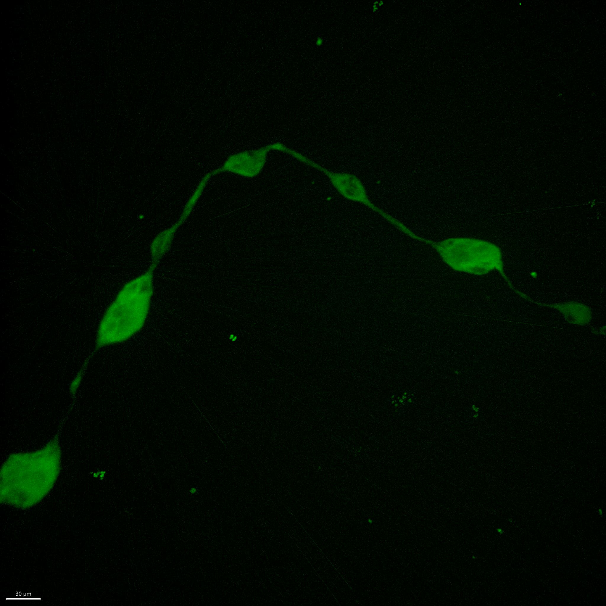
Axon running inside a human brain organoid.
Axon running inside a human brain organoid.
At least I think this is axon, as the structure we observed shows classical axonal swellings normally considered pathological in neurodegenerative models.
🟩Green fluorescent protein 🟪Glial Fibrillary Acidic Protein (GFAP)
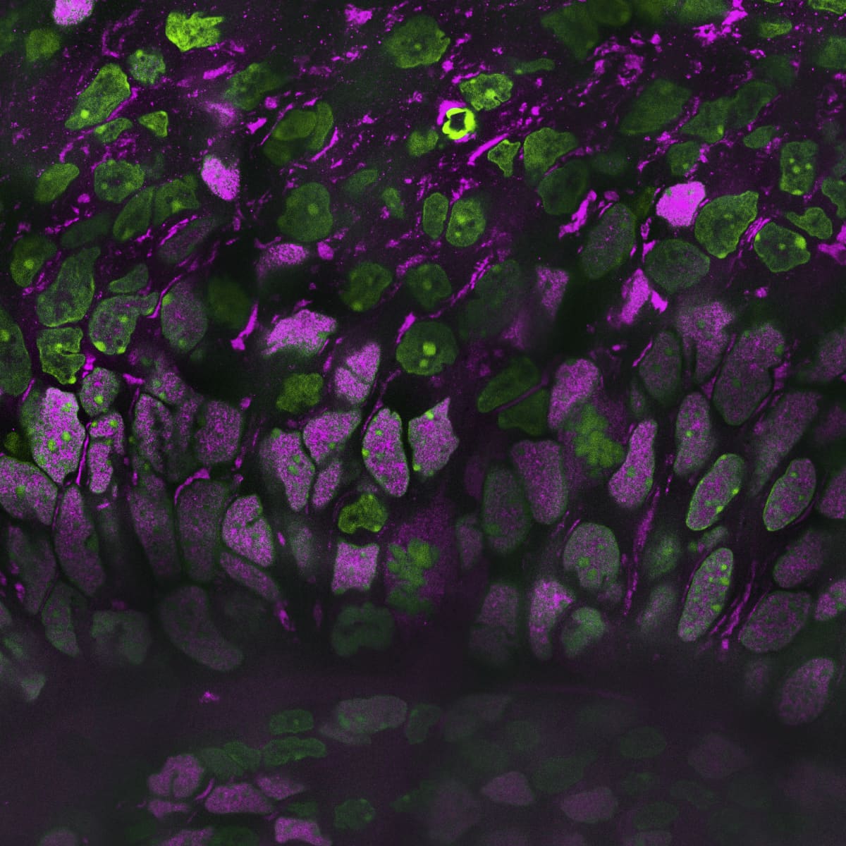
Ventricular zone in a human brain organoid.
Ventricular zone in a human brain organoid.
The dark arch along the bottom is the ventricular zone, a cavity that appears when stem cells arrange themselves into rings, exactly as they do in a real developing brain.
The cells hugging that wall are the earliest stem cells of the lot, and they're the ones that divide and turn into neurons. You can spot them by 🟪, a protein that switches on in brain stem cells before they commit to becoming anything in particular.
🟩 DNA 🟪 SOX2 — the badge of a brain stem cell
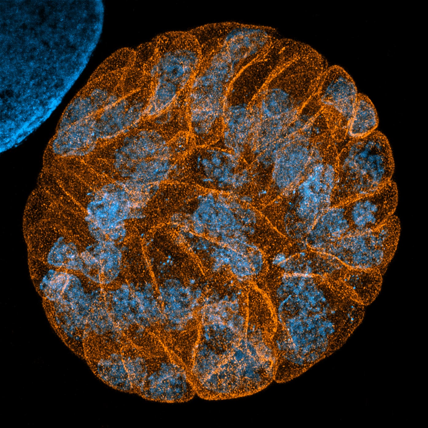
Chronic-stage Toxoplasma cyst with parasites inside
Winner — American Society for Microbiology Journal 2024
Toxoplasma is a parasite that settles into the brain for life, sealed inside a tough little pod called a cyst.
I wanted to look inside one using expansion microscopy — a trick where you swell the whole sample in a water-absorbing gel until its tiniest structures are big enough for an ordinary microscope to see.
Nobody had published this on cysts, and I soon found out why: the cyst wall shrugs off the chemicals that make everything else expand. It took a while to crack, but it worked — and now any lab can do it, no super-resolution microscope required.
🟧 Cyst wall 🟦 DNA
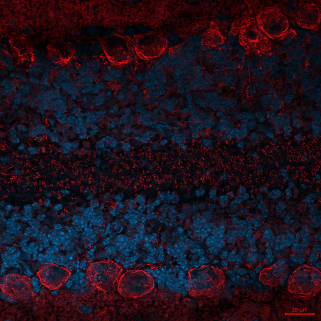
Mouse Purkinje neurons
The red rings at the top and bottom are Purkinje neurons, the cells that keep you balanced and coordinated — normally famous for their sprawling branches, but here recognisable by their plump cell bodies, lined up single-file along the folds of the cerebellum. The two thick blue bands between them are the tightly packed nuclei of granule cells, the smallest and most numerous neurons in the brain. The red glow itself comes from Kv3.3, a protein that acts like a fast shutter on the cell membrane, cutting each electrical pulse short so the neuron can fire again immediately. When the gene for Kv3.3 is mutated, Purkinje cells sicken and die, causing spinocerebellar ataxia type 13 — a rare inherited disease that gradually robs people of balance, walking, speech and eye control.
🟥 Purkinje neurons (Kv3.3) 🟨 Synapses (Bassoon) 🟦 DNA
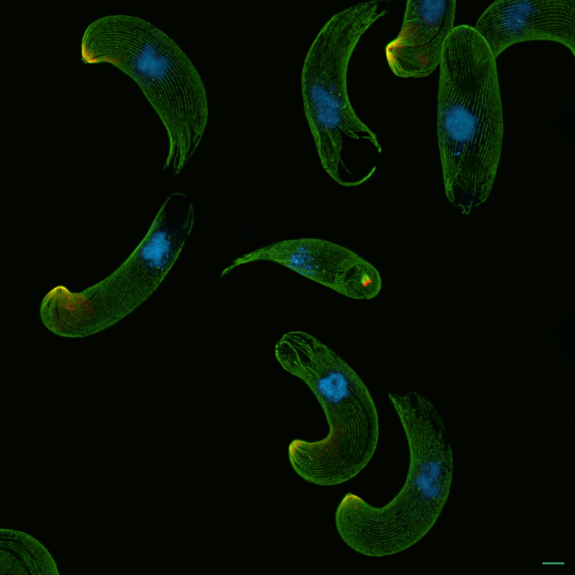
Malaria parasite that forms in mosquito midgut
Malaria, caught mid-journey.
These are ookinetes — the form the parasite takes inside a mosquito's stomach, after mating there. Their one job is to drill through the gut wall and settle in, eventually sending a new generation up to the mosquito's salivary glands, loaded and ready for the next bite.
Look for the "floating" green ring at the parasite's tip: that's the tool it breaks in the host cell with, and essentially its mouth.
🟩 Parasite skeleton 🟥 Structures at the parasite tip 🟦 DNA
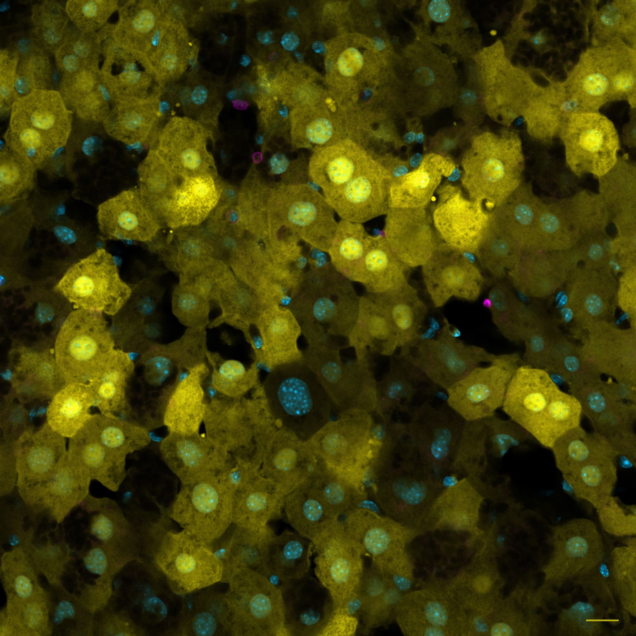
Mouse liver, under patrol of immune cells
A mouse liver whose cells have been made to glow. The yellow comes from a fluorescent protein delivered into them by a harmless engineered virus — a standard way of getting cells to announce themselves with light. Scattered among the big liver cells are a few much smaller ones in magenta: T cells, the immune system's patrol, going about their rounds.
🟨 Liver cells, glowing with Venus (a yellow fluorescent protein) 🟪 Immune cells (T cells, CD3) 🟦 DNA
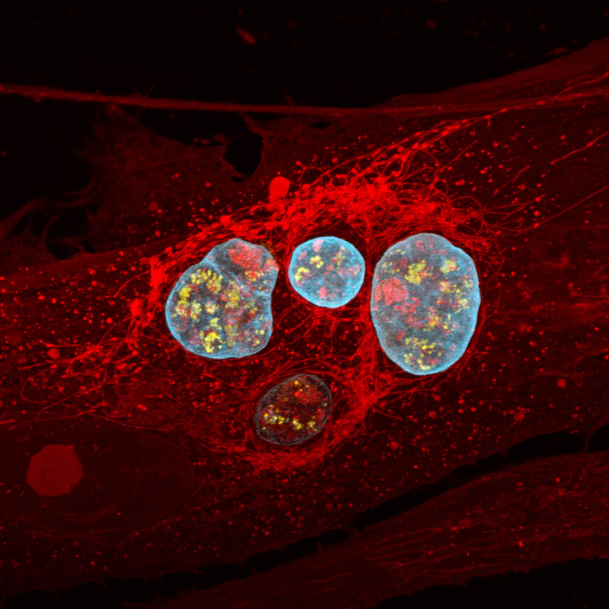
Chronic Toxoplasma infection of human skin cells
Toxoplasma is a parasite that moves in for good: once the infection turns chronic, it walls itself into cysts that no drug can reach. These ones are sitting inside human skin cells.
Look for the red net around each cyst — that's the host cell crowding its own power plants (mitochondria) up against the intruder, a siege of sorts.
🟦 Cyst wall (GAP45) 🟥 All proteins (NHS-ester) 🟨 A parasite protein that keeps the cyst interior in order (GRA2)
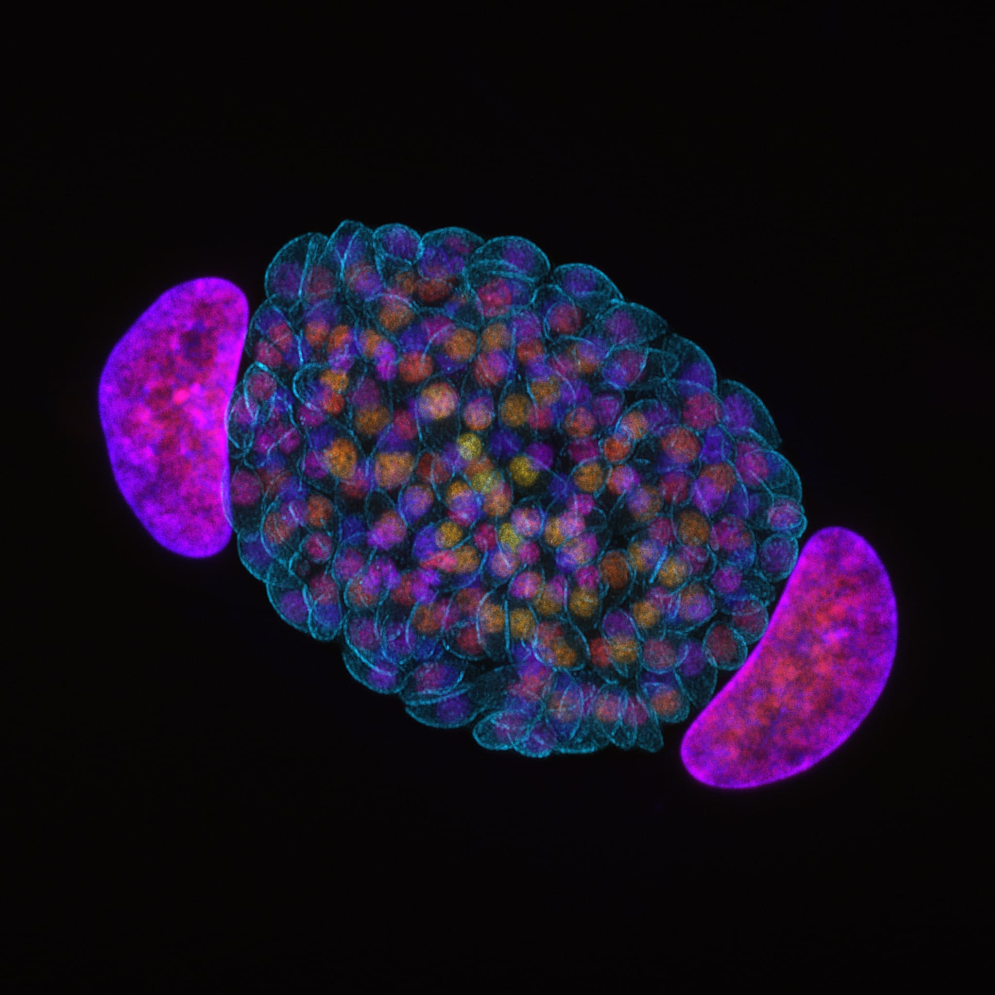
Toxoplasma parasites in a human skin cell
Toxoplasma parasites in day three of acute infection, sitting snugly between two nuclei of a human skin cell.
Toxoplasma is documented to dysregulate the host cell cycle - host cells like this fibroblast could duplicate their DNA but never finish the division, remaining binucleate.
🟦Toxoplasma inner shell 🟨🟧🟥🟪 DNA (depth colour-coded)


